
What Are Tumor Markers? Origins, Types, Positive Rates by Cancer, Cost, and How to Read Results, Checked Against Society Guidelines and Studies

PR This article contains affiliate links to Rakuten Ichiba (“See on Rakuten”). What is written here is based on materials from public agencies and medical societies and on published studies, whether or not there is an ad. The “See on Amazon.co.jp” and “See on Yahoo! Shopping” links are also affiliate links.
About this article This article organizes how the tumor marker test works and what is written in materials from medical societies and public agencies. It does not judge what any one person’s test result means. If a result worries you, talk to the medical institution that did the test or to your regular doctor.
Your health checkup or full-day medical exam (ningen dock) results list items like “CEA,” “CA19-9,” and “PSA,” and yours is a little above the reference value. Or you are signing up for a ningen dock and wondering whether to add the optional “tumor marker” tests. When you hear “a blood test that can detect cancer,” getting it sounds reassuring.
This article checks the following against the guidelines of the Japanese Society of Laboratory Medicine, the National Cancer Center Japan, materials from public agencies overseas, and published studies.
- What tumor markers are, and when and by whom they were discovered
- What types there are, and what substance in the body each one measures
- Which marker is positive in which cancer, and about what percent of the time (by cancer type and stage)
- When a checkup result is “positive,” what share of those people really have cancer
- What raises markers without cancer, and why markers can stay normal when cancer is present
- How to read results and the criteria used, and the cost (insurance and self-pay)
- How Japan and other countries differ in their ratings of these tests as screening
Conclusions
- Tumor markers are substances such as proteins that cancer cells make in larger amounts. Most are also made by normal cells, so they can rise from non-cancer diseases, smoking, pregnancy, menstruation, reduced kidney function, and medications (National Cancer Center Japan, Japanese Society of Laboratory Medicine)
- The story begins with the Bence Jones protein in 1847. In the 1960s came the fetal proteins AFP and CEA, and after the invention of monoclonal antibodies in 1975 came CA19-9, CA125, and others. Many, such as SCC, ProGRP, and AFP-L3, came out of Japanese research
- The share of people with cancer who test positive (positive rate, or sensitivity) is about 78% for CA19-9 in pancreatic cancer, about 80% for PSA in prostate cancer, about 50% for CEA in colorectal cancer, and about 24% for CEA in stomach cancer (approximate values from JSLM2021 of the Japanese Society of Laboratory Medicine). But the earlier the cancer, the lower the rate: CA19-9 in stage I stomach cancer was 3%
- In screening people with no symptoms, the rates are even lower. In National Cancer Center Japan checkup data, CEA was positive in only 7.8% of the stomach, duodenal, and colorectal cancers that were found, and of the people who were CEA-positive, only 3.7% actually had cancer
- Tumor markers are not part of the country’s five cancer screenings, and the National Cancer Center Japan says that “an effect as cancer screening … has not been shown.” PSA alone is rated differently by different groups: the National Cancer Center Japan does not recommend it for municipal screening, while the Japanese Urological Association strongly recommends it. Overseas, ovarian cancer screening with CA125 did not reduce deaths in a large trial
- On cost, insurance covers these tests only “when cancer is strongly suspected,” and at 30% copay the testing costs about 850 yen (1 item) to about 1,710 yen (4 or more items). Ningen dock options (self-pay) run roughly 2,000 to 3,000 yen and up per single test. Reference values differ by hospital and reagent, so the societies write that results should be judged by the size of the value, its trend over time, and other tests taken together
Items Introduced in This Article
Where they fit the topic, the article introduces the following books (Rakuten Ichiba links).
| For people who | Item introduced | Why it was chosen (fact) | Where |
|---|---|---|---|
| Want to look up what each lab test means | 『パッと引けてしっかり使える 検査値の読み方ポケット事典[第5版]』 (Seibido Shuppan) | Published 2023. The table of contents has “Chapter 7: Tumor Marker Tests” | 結果はどうみる? |
| Want to know how to read checkup results as a whole | 『健診結果の読み方 気にしたほうがいい数値、気にしなくていい項目』 (Kodansha+α Shinsho) | Published 2024. The table of contents has “Chapter 10: Cancer Screening” | 国が勧めるがん検診との関係 |
What Are Tumor Markers?
The Cancer Information Service of the National Cancer Center Japan describes tumor markers as follows.
Tumor markers are substances, mainly proteins, made mostly by cancer cells, and they have features that differ by type of cancer and by organ. (National Cancer Center Japan, Cancer Information Service, “Tumor Marker Tests,” updated July 8, 2024)
The Japanese Society of Laboratory Medicine’s “Clinical Laboratory Guidelines JSLM2021” defines a tumor marker test as “a test that measures serum concentrations of substances produced by tumor cells, or substances the body produces in response to a tumor,” and gives its purposes as aiding the diagnosis of a tumor, following the effect of treatment over time, and monitoring for recurrence.
The key point is that most tumor markers are not substances made only by cancer. The U.S. National Cancer Institute (NCI) explains that many tumor markers are “proteins made by both normal cells and cancer cells, with cancer cells making more,” and lists three limits (NCI “Tumor Markers” fact sheet, December 7, 2023).
- A high tumor marker does not necessarily mean there is cancer
- Diseases other than cancer can also raise tumor markers
- Not everyone with that cancer has a raised tumor marker
And JSLM2021 states plainly:
Tumor markers do not show a significant increase in early cancer, so they are not suited to screening for early detection of cancer. (Japanese Society of Laboratory Medicine, “Clinical Laboratory Guidelines JSLM2021,” Tumor Marker Tests and Companion Diagnostic Tests, p. 87)
The same passage names the PSA for prostate cancer as an exception. Later in this article we look at why PSA is treated differently, and at where Japan and other countries disagree about it.
The Origins of Tumor Markers
The “first tumor marker” was a urine test in 1847
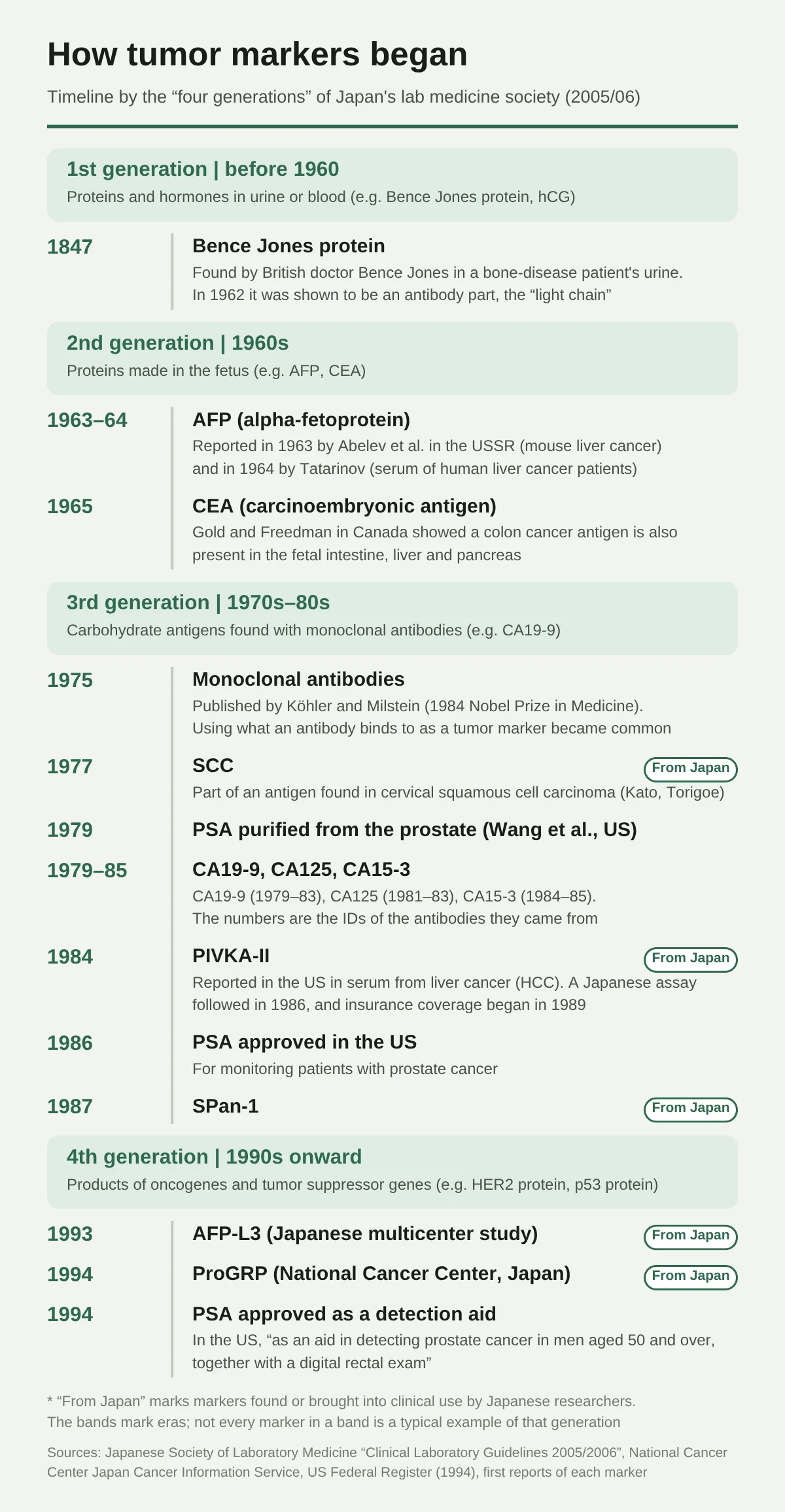
What is often called the “first tumor marker” is a substance (the Bence Jones protein) that the British physician Henry Bence Jones found in 1847 in the urine of a patient with a bone disease. It was reported in 1848 in a Royal Society journal. It was identified as the “light chain,” a part of an immunoglobulin (antibody), more than 100 years later, in 1962 (Edelman and Gally, 1962). The Bence Jones protein is what appears in large amounts in the urine of people with what we now call multiple myeloma (Sewpersad and Pillay 2021). Some sources give 1846 as the year of discovery.
The 1960s: fetal proteins reappear in cancer
Research on tumor markers got going in earnest in the 1960s. What was found was that proteins made in the fetus, and no longer made by the time of birth, show up in the blood again when cancer develops.
- AFP (alpha-fetoprotein): Abelev and colleagues in the Soviet Union reported it in mouse liver cancer in 1963, and Tatarinov reported it in the serum of human liver cancer patients in 1964. AFP is a protein made in the fetal liver and yolk sac
- CEA (carcinoembryonic antigen): In 1965, Gold and Freedman in Canada showed that an antigen found in human colorectal cancer is also present in the intestine, liver, and pancreas of fetuses 2 to 6 months into pregnancy, and named it “carcinoembryonic antigen”
1975: The era of searching for “targets” with monoclonal antibodies
In 1975, Köhler and Milstein announced a method of producing one specific type of antibody indefinitely (monoclonal antibodies). This work led to the 1984 Nobel Prize in Physiology or Medicine.
After this, the approach spread of making antibodies using cancer cells and using whatever those antibodies bind to as a tumor marker. Examples are CA19-9 (1979–1983, Koprowski and colleagues), CA125 (1981–1983, Bast and colleagues), and CA15-3 (1984–1985). The numbers in the names (19-9, 125, and so on) come from the numbers of the antibodies they were based on.
Many markers were also discovered, or brought into clinical use, by Japanese researchers.
- SCC: A portion of an antigen called TA-4, which Kato and Torigoe of Japan found in squamous cell cancer of the uterine cervix in 1977
- PSA: “Gamma-seminoprotein,” a semen protein reported in 1971 by Hara and colleagues of Japan, was later found to be the same substance as PSA (confirmed in a review article; the 1971 original paper was not checked). PSA was purified from the prostate in 1979 by Wang and colleagues in the U.S.
- PIVKA-II: It was reported in the U.S. in 1984 to be found in the serum of 91% of hepatocellular carcinoma patients (Liebman and colleagues), and in 1986 an assay using monoclonal antibodies was reported in Japan. In Japan it became measurable under insurance in 1989
- AFP-L3 (1993, a multicenter study in Japan), ProGRP (1994, the National Cancer Center Research Institute), and SPan-1 (1987) are also from Japanese research
The history of PSA’s approval
In the U.S. in 1986, a PSA test kit was approved for monitoring patients with prostate cancer. It was approved as “an aid, together with a digital rectal exam, in detecting prostate cancer in men 50 and older” only in 1994 (Federal Register). It started out as a test for following people already known to have cancer.
The Japanese Society of Laboratory Medicine’s “four generations”
The Japanese Society of Laboratory Medicine’s guideline (2005/2006 edition) divides tumor markers into four generations by when they were found.
| Generation | Era | Representative examples | Features |
|---|---|---|---|
| First generation | Before 1960 | Bence Jones protein, hCG | Proteins and hormones in urine and blood |
| Second generation | 1960s | AFP, CEA | Proteins made during fetal life |
| Third generation | 1970s–80s | CA19-9 and others | Carbohydrate antigens found with monoclonal antibodies |
| Fourth generation | 1990s onward | HER2 protein, p53 protein | Products of oncogenes and tumor suppressor genes |
Types of Tumor Markers
The Cancer Information Service of the National Cancer Center Japan lists the following tumor markers by organ.
| Organ | Main tumor markers |
|---|---|
| Lung | CYFRA, CEA, ProGRP, NSE |
| Stomach, colon | CEA, CA19-9 |
| Liver | AFP, PIVKA-II, AFP-L3 fraction |
| Pancreas | CA19-9, SPan-1, DUPAN-2, CEA, CA50 |
| Prostate | PSA |
| Breast | CEA, CA15-3 |
| Ovary | CA125 |
Source: The list in the National Cancer Center Japan Cancer Information Service “Tumor Marker Tests” (updated July 8, 2024), turned into a table.
One marker does not point to only one cancer. For example, CEA rises not only in colorectal cancer but also in lung, stomach, and breast cancer. Conversely, there are several markers for a single cancer, and doctors choose a combination to suit the type of cancer and the purpose.
A List of Tumor Markers by Substance
Tumor markers become easier to follow when you sort them by the kind of “substance” they measure. What they are varies: glycoproteins (proteins with sugars attached), carbohydrate antigens (which use the shape of a sugar chain as the target), enzymes, hormones, and fragments of the cell’s scaffolding.
| Marker | Kind of substance (what it is) | Where it is normally made | Cancers mainly watched |
|---|---|---|---|
| CEA (carcinoembryonic antigen) | Glycoprotein | Fetal intestine, liver, and pancreas. In adults, cells of the lining of the colon and elsewhere | Digestive cancers such as colorectal cancer; lung cancer |
| AFP (alpha-fetoprotein) | Glycoprotein | Fetal liver and yolk sac | Hepatocellular carcinoma, germ cell tumors |
| AFP-L3 fraction | AFP with a sugar called fucose attached at its base | The share rises in the AFP made by hepatocellular carcinoma | Hepatocellular carcinoma |
| PIVKA-II | Abnormal prothrombin (an incomplete blood-clotting protein made when vitamin K is lacking) | Liver (normal prothrombin is a clotting factor made in the liver) | Hepatocellular carcinoma |
| CA19-9 | Carbohydrate antigen (a sugar chain called sialyl Lewis A). In the blood it rides mainly on mucin (a mucus protein) | Mucus of the salivary glands, pancreatic ducts, and so on | Pancreatic cancer, biliary tract cancer, stomach and colorectal cancer |
| CA125 | Part of a mucin (MUC16) | Lining of the fallopian tubes and uterus; the pleural and peritoneal membranes | Ovarian cancer |
| CA15-3 | Part of a mucin (MUC1) | Secretory cells of the mammary gland | Breast cancer (mainly for following metastasis and recurrence) |
| PSA (prostate-specific antigen) | Enzyme (a serine protease that breaks down proteins) | Prostate. Works to thin out semen | Prostate cancer |
| SCC | A protein that stops protein-digesting enzymes (a serpin) | Squamous epithelium of the skin and mucous membranes | Cervical cancer; squamous cell cancers of the lung, esophagus, and head and neck |
| CYFRA | Fragment of the cell scaffold (cytokeratin 19) | Scaffold of the cells of the mucous membranes | Lung cancer (especially squamous cell carcinoma) |
| ProGRP | A peptide that is the precursor of a hormone (gastrin-releasing peptide) | Released in large amounts by small cell lung cancer cells | Small cell lung cancer |
| NSE (neuron-specific enolase) | Enzyme (the neuronal form of an enzyme that breaks down sugar) | Nerve cells and neuroendocrine cells | Small cell lung cancer, neuroblastoma |
| DUPAN-2 | Carbohydrate antigen (sialyl Lewis C, a form one step before CA19-9) | Lining of the pancreatic ducts | Pancreatic cancer |
| SPan-1 | Carbohydrate antigen (reacts with both sialyl Lewis A and sialyl Lewis C) | Mucus of salivary glands and elsewhere | Pancreatic cancer |
| CA72-4 | Sugar chain of a mucin-like glycoprotein (TAG-72) | Almost none in normal tissue | Stomach cancer, ovarian cancer |
| STN (sialyl Tn) | Carbohydrate antigen (the base of a mucin-type sugar chain) | Almost none in normal tissue | Ovarian cancer (especially mucinous), digestive cancers |
| NCC-ST-439 | Carbohydrate antigen (sialyl Lewis X) | A little in salivary glands and the trachea | Breast, stomach, and colorectal cancer |
| Elastase 1 | Digestive enzyme of the pancreas | Pancreas | Pancreatic cancer (also rises in pancreatitis) |
| sIL-2R (soluble interleukin-2 receptor) | A receptor of immune cells that has been released into the blood | Activated T lymphocytes | Malignant lymphoma, adult T-cell leukemia |
| hCG | Hormone (a glycoprotein hormone) | Placenta (rises in pregnancy) | Choriocarcinoma, germ cell tumors |
| Anti-p53 antibody | An antibody made by one’s own body (against the p53 protein altered in cancer cells) | Immune cells | Esophageal, colorectal, and breast cancer |
| HE4 | Protein (shaped like proteins that block proteases) | Lining of the epididymis | Ovarian cancer |
Source: Compiled from the original papers of Gold and Freedman 1965, Magnani et al. 1983, Yin and Lloyd 2001, Liebman et al. 1984, Schneider et al. 1995, Pujol et al. 1993, Miyake et al. 1994, Kawa et al. 1994, Kirchhoff et al. 1991, and others, the guidelines of the Japanese Society of Laboratory Medicine (2005/2006 and 2021), and the NCI list.
Looking at the table, you can see that the where it is normally made column is full of places that are not cancer. Tumor markers are mostly not “substances only cancer makes” but “substances the body already has, which tend to increase when cancer is present.” This is one reason values can rise without cancer.
Which Marker Is Positive in Which Cancer, and What Percent of the Time?
Positive rates by type of cancer
The next figure shows the “approximate sensitivity” in Table 1 of JSLM2021 from the Japanese Society of Laboratory Medicine. Sensitivity (positive rate) means the share of people who have that cancer whose tumor marker is above the reference value.
For example, it means that about 78% of people with pancreatic cancer have CA19-9 above the reference value (37 U/mL), and about 80% of people with prostate cancer have PSA above the reference value (4 ng/mL).
However, JSLM2021 itself cautions about these numbers as follows.
Even in studies of the same marker in the same tumor, the positive rate differs greatly depending on the test method, the cutoff value set, and the distribution of stages in the patient group studied, so sensitivity is not fixed at a specific number.
Here are some reported examples for markers not in the figure. In each, the population and the reference value differ from paper to paper.
| Marker | Cancer | Reported positive rate | Source |
|---|---|---|---|
| CA19-9 | Pancreatic cancer | 79% (70 to 90% depending on the study) | Goonetilleke and Siriwardena 2007 (systematic review) |
| CEA | Stomach cancer | 21.1% | Shimada et al. 2014 (task force of the Japanese Gastric Cancer Association) |
| CA19-9 | Stomach cancer | 27.8% | Shimada et al. 2014 |
| CA72-4 | Stomach cancer | 30.0% | Shimada et al. 2014 |
| DUPAN-2 | Pancreatic cancer | 63% | Gong et al. 2025 (meta-analysis) |
| SPan-1 | Pancreatic cancer | 72.0% (64 people) | Takeda et al. 1991 |
The earlier the cancer, the less likely a tumor marker is to rise
Positive rates change greatly with how far the cancer has advanced (stage). The next figure shows positive rates by stage from the 2005/2006 guideline of the Japanese Society of Laboratory Medicine.
For CA19-9, the rate in stomach cancer is 3% at stage I and 67% at stage IV. For PIVKA-II in hepatocellular carcinoma, it goes from 0% at stage I to 100% at stage IV. Figures such as “about 80%” in Figure 1 are overall numbers that include people with advanced cancer. The earlier the stage at which you want to find the cancer, the less likely a tumor marker is to rise. This is why the Japanese Society of Laboratory Medicine writes that they are “not suited to screening for early detection.”
If It’s Positive, Is It Cancer? “Positive Rate” and “Probability of Having Cancer” Are Different
Results for 12,349 people who had CEA measured at a checkup
The “positive rate” in Figure 1 was the percent who test positive among people with cancer. What people having a checkup want to know is the reverse: among people who tested positive, what percent really have cancer (the positive predictive value). These two are completely different numbers.
There is data from people without symptoms who had voluntary cancer screening (screening like a ningen dock) at the screening center of the National Cancer Center Hospital in Japan. In this study, upper and lower endoscopy was used to confirm whether they had stomach, duodenal, or colorectal cancer (upper and lower gastrointestinal cancer, in the paper’s grouping), and the performance of CEA and CA19-9 was examined (Sekiguchi and Matsuda, Scientific Reports 2020).
- Of the 230 people in whom stomach, duodenal, or colorectal cancer was found, CEA was above the reference value in 18 (7.8%). The other 212 were within the reference value
- Of the 487 people whose CEA was above the reference value, 18 (3.7%) actually had these cancers
- For CA19-9 (reference value 37 U/mL), sensitivity was 7.4% and positive predictive value was 2.7%
- Among the 7,616 people who also had whole-body imaging, even counting cancers anywhere in the body, the positive predictive value was 4.1% for CEA and 5.8% for CA19-9
- For stomach, duodenal, and colorectal cancer (analysis of 12,349 people), raising the reference values to CEA 10 ng/mL and CA19-9 74 U/mL raised the positive predictive value to 11.8% and 4.9%, but lowered sensitivity to 2.6% and 1.7%
In other words, even a marker that is positive in 70 to 80% of people at a cancer hospital, when used for screening people without symptoms, was positive in less than 10% of those who had cancer, and more than 90% of those who tested positive did not have cancer. The title of the paper was also “CEA and CA19-9 have limited usefulness for screening for gastrointestinal and whole-body cancers.”
What happened to people with high CA19-9 at a ningen dock
Reports from Japanese ningen docks show nearly the same numbers.
| Population | People with high CA19-9 | Of those, people with cancer | Source |
|---|---|---|---|
| Screening center of the Cancer Institute Hospital of JFCR (Ariake), 32,508 exams in total | 790 exams (about 2.4%) | 8 of the 320 people who were retested (2.5%) | Suzuki et al. 2015, Ningen Dock 30:22-29 |
| Ningen dock, 15,773 people (fiscal 2010) | 308 people (2.0%) | 10 of the 243 who had a second exam (4.1%). Pancreatic cancer in 4 | Yajima et al. 2014, Ningen Dock 29:610-615 |
Alongside the low positive predictive value, the report by Yajima et al. also notes that for 5 malignant tumors (including 3 pancreatic cancers) that were found because of an abnormal CA19-9, treatment with complete surgical removal (curative resection) was possible. The authors followed CA19-9 over time every 3 months. Most of the people who tested positive did not have cancer, yet some of the cancers that were found could be treated early. Both facts are written in the same report.
Why is it so low?
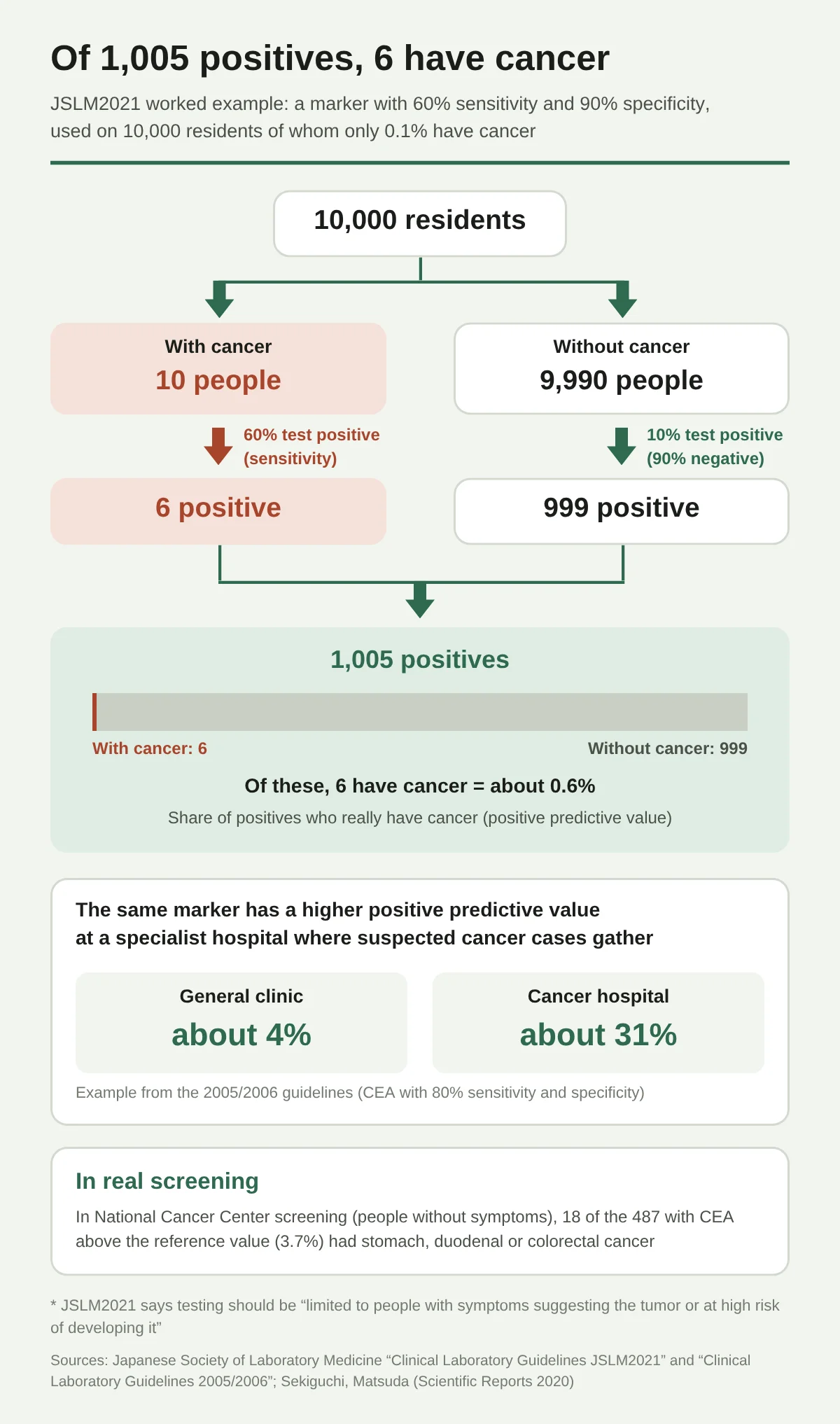
The reason is that, among the people who have a checkup, very few have cancer. JSLM2021 gives a calculation example. If you use a marker with 60% sensitivity and 90% specificity (90% of people without cancer test negative) on 10,000 residents of whom only 0.1% have cancer:
- Of the 10 people with cancer, 6 test positive
- Of the 9,990 people without cancer, 10%, or 999, test positive
- Of the 1,005 positives, 6 have cancer, which is about 0.6%
The same marker has a higher positive predictive value when it is used in a specialist hospital where people suspected of having cancer gather (the 2005/2006 guideline gives an example for CEA with 80% sensitivity and specificity: about 4% at a general clinic and about 31% at a cancer hospital). This is why JSLM2021 writes that “testing needs to be limited to people with symptoms suggesting that tumor or people at high risk of developing it.”
Raised Without Cancer, Not Raised Despite Cancer
Causes of a rise other than cancer
Tumor markers also rise with diseases and body conditions other than cancer. The main ones are summarized from the guidelines of the Japanese Society of Laboratory Medicine and from studies.
| Marker | Main non-cancer causes of a rise | Example numbers |
|---|---|---|
| CEA | Smoking, aging, hepatitis and cirrhosis, inflammatory bowel disease, pancreatitis, diabetes | In voluntary screening, CEA was positive in 12.5% of smokers and 2.8% of nonsmokers (Sekiguchi 2020) |
| CA19-9 | Gallstones, cholangitis, obstructive jaundice, pancreatitis, endometriosis, poorly controlled diabetes | About 20% of people with gallstones are positive (guideline 2005/2006) |
| CA125 | Endometriosis, menstruation, pregnancy, peritonitis, pleurisy, pancreatitis, cirrhosis | - |
| PSA | Benign prostatic hyperplasia, prostatitis, urinary retention, procedures on the urethra, ejaculation | Still raised 24 hours after ejaculation in 40% of people (a study of 20 people, Herschman 1997) |
| AFP | Chronic hepatitis, cirrhosis, pregnancy | 20 to 40% of people with chronic hepatitis or cirrhosis are positive (guideline 2005/2006) |
| PIVKA-II | Vitamin K deficiency, warfarin (a blood thinner), some antibiotics and anti-tuberculosis drugs, obstructive jaundice | - |
| SCC | Skin diseases (atopic dermatitis, pemphigus, and others), pneumonia and bronchitis, renal failure | 95.1% of people with renal failure before dialysis are positive (Odagiri 1991) |
| CYFRA | Hepatitis and cirrhosis, lung inflammation, reduced kidney function | 57% of people on dialysis are above the reference value (Nakahama 1998) |
| ProGRP | Reduced kidney function | 90% of people on dialysis are above the reference value (Nakahama 1998) |
| NSE | Cerebrovascular disease, encephalitis, hemolysis (destruction of red blood cells after the blood draw) | - |
Source: JSLM2021 of the Japanese Society of Laboratory Medicine, Table 1 and the chapter on liver cancer; the same society’s 2005/2006 edition; Sekiguchi and Matsuda 2020; Herschman et al. 1997; Odagiri et al. 1991; Nakahama et al. 1998.
There is also talk that long bicycle rides raise PSA, but a study found a rise (Mejak et al. 2013, an average increase of 9.5%) while a study pooling 8 studies found no meaningful difference (Jiandani et al. 2015), so the conclusions differ.
Why it may not rise even when there is cancer
- Early cancer: As Figure 2 above shows, the positive rate falls sharply at stage I
- Some people cannot make CA19-9 from birth: CA19-9 is a sugar chain made through the Lewis blood group system. People who are “negative” in the Lewis system (about 10% of Japanese) show almost no rise in CA19-9 even with pancreatic cancer (guideline 2005/2006). Of 2,418 people who had pancreatic cancer surgery in Japan, 7.7% were people who do not make CA19-9 (Omiya et al. 2026). In such people DUPAN-2 can sometimes be useful
- Lowered by medication: People taking 5-alpha reductase inhibitors, used to treat benign prostatic hyperplasia and male pattern hair loss, have PSA that falls by about half on average (Japanese Urological Association, “Guidelines for Prostate Cancer Screening, 2018 edition”)
Criteria for Judging Results
A guide to reference values (cutoff values)
Tumor marker results are often sorted into positive and negative by “whether the value is above the reference value (cutoff).” Below are the values in Table 1 of JSLM2021 of the Japanese Society of Laboratory Medicine, alongside examples of hospital reference values.
| Marker | Cutoff value in JSLM2021 Table 1 | Example of a hospital reference value |
|---|---|---|
| CEA | 5.0 ng/mL | Aichi Cancer Center: 5.0 or less |
| AFP | 15 ng/mL | Aichi Cancer Center: less than 10.0 |
| CA19-9 | 37 U/mL | Aichi Cancer Center: less than 37.0; Wakayama Medical University: 37 or less |
| CA125 | 35 U/mL | - |
| CA15-3 | 27 U/mL | Aichi Cancer Center: 25.0 or less |
| PSA | 4 ng/mL | By age: ages 50-64: 3.0, ages 65-69: 3.5, age 70 and over: 4.0 (Japanese Urological Association, 2018 edition) |
| PIVKA-II | 40 mAU/mL | Wakayama Medical University: 33.3 mAU/mL or less |
| SCC | 1.5 ng/mL | Aichi Cancer Center: 2.5 or less |
| CYFRA | 3.5 ng/mL | Aichi Cancer Center: 3.5 or less |
| NSE | 10 ng/mL | - |
| ProGRP | (not given in the table) | Aichi Cancer Center: less than 81.0 pg/mL |
| Anti-p53 antibody | 1.3 U/mL | - |
Source: JSLM2021 of the Japanese Society of Laboratory Medicine, Table 1 (p. 88); Aichi Cancer Center, “List of Clinical Laboratory Reference Values” (in effect from February 1, 2026); the laboratory guide of Wakayama Medical University Hospital; Japanese Urological Association, “Guidelines for Prostate Cancer Screening, 2018 edition.”
Even for the same AFP, the values are 15 and 10, and for SCC 1.5 and 2.5, so reference values differ by hospital and by measurement kit. Wakayama Medical University says its reference values are based on the package insert of the reagent it uses. A study of international external quality assessment data reported that for CA125, CA15-3, and CA19-9 the differences between measurement methods were large and did not meet minimum standards (van Rossum, Clinical Chemistry 2024; checked from the abstract and a society summary). This is why it is better not to line up values measured at different hospitals and compare them as they are.
The “A to E rating” of a ningen dock and tumor markers
The Japan Society of Ningen Dock and Preventive Medicine sets rating categories that sort results into A (no abnormality), B (mild abnormality), C (retest or lifestyle improvement needed), D (detailed exam or treatment needed), and E (under treatment). However, tumor markers are in neither the fiscal 2026 rating table nor the basic test items. The rating given to tumor markers on a ningen dock result sheet follows criteria set by that facility.
Age-specific reference values are also used for PSA
The Japanese Urological Association’s “Guidelines for Prostate Cancer Screening, 2018 edition” gives the reference value for PSA as “4.0 ng/mL, or by age group (ages 50-64: 3.0, ages 65-69: 3.5, age 70 and over: 4.0),” and shows the following flow.
| PSA value | Next step shown in the 2018 guideline |
|---|---|
| 1.0 ng/mL or less | Screen again in 3 years |
| 1.1 ng/mL up to the reference value | Screen again in 1 year |
| Above the reference value | Refer to a urology specialist who can do a prostate biopsy |
The same society’s “2025 edition” came out in September 2025, but this article has not been able to check its text.
When biopsies were done in the PSA “gray zone” of 4.1 to 10 ng/mL, the share in which cancer was found ranged in Japanese reports from 23.6% (Hamamatsu Rosai Hospital, Kobayashi et al. 2004) to 46% (a method that increased the number of biopsy cores, Ito et al. 2002). Conversely, in a large U.S. trial, when 2,950 men whose PSA never exceeded 4.0 had biopsies, prostate cancer was found in 15.2% (Thompson et al., NEJM 2004).
How to Read the Results
“A little over” and “very high” are different
JSLM2021 of the Japanese Society of Laboratory Medicine writes the following about how to read results (pp. 87-88).
In practice, judgment is not made on whether the marker is positive or negative alone; clinical judgment differs depending on the size of the value, that is, whether it is positive by only slightly exceeding the cutoff value or abnormally high.
The substances that serve as tumor markers are not made only by tumor cells but also by normal cells, and some tumor markers rise with inflammation, aging, pregnancy, reduced kidney function, medications, and so on, so interpreting the results requires a comprehensive judgment that takes into account symptoms, physical findings, and the results of other tests.
The same document says that when AFP or PSA is only slightly raised, it is also useful to check the more specific AFP-L3 fraction or the free PSA/total PSA ratio, to combine several markers, and to follow the trend over time.
What can be read from this, kept within the wording of the guideline, is as follows.
| What to check | Why | Basis |
|---|---|---|
| How far above the reference value | Judgment differs depending on whether it is slightly above or very high | JSLM2021 |
| How it moved compared with previous values | Following the trend is considered useful. Values from another hospital may use different reference values and methods | JSLM2021, van Rossum 2024 |
| Whether there is a cause of a rise other than cancer | Smoking, aging, pregnancy, menstruation, reduced kidney function, medications, and benign diseases can also raise it | JSLM2021 Table 1 |
| Symptoms and the results of other tests | It cannot be decided from tumor markers alone; it is judged comprehensively | JSLM2021 |
Within what I looked at, I did not find a procedure set by a medical society or public agency, written for the general public, that says which tests to have in which order when a tumor marker is high. For PSA, there is the flow from the Urological Association in the previous section. If your result sheet says “detailed exam needed,” follow the instructions of the checkup facility and talk with a medical institution.
A “tumor marker retest” does not count as a detailed exam after cancer screening
If a person is told “detailed exam needed” in a municipal cancer screening (stomach, colorectal, lung, breast, cervical) and has only a tumor marker measured instead of the detailed exam, that is not treated as a detailed exam. A document of the Ministry of Health, Labour and Welfare (MHLW) (“On the Cancer Screening Program,” July 2024) states in its reporting instructions: “Cases in which a test inappropriate as a detailed exam (a repeat fecal occult blood test, CT, tumor markers, and so on) was done are to be counted as not having had the exam.”
The Cost of Tumor Markers
Insurance covers them only “when cancer is strongly suspected”
Under the rules of the medical fee schedule, health insurance covers tumor marker tests only for “patients strongly suspected of having a malignant tumor from the results of the examination and of tests other than tumor markers.” As a rule it is once, until the diagnosis is confirmed. When a person without symptoms has it measured as a checkup, insurance cannot be used and it is self-pay (health checkups are outside insurance-covered care).
As exceptions, AFP and PIVKA-II in people with cirrhosis or chronic hepatitis B or C (up to once a month), CA125 in endometriosis, and others can be measured under insurance under set conditions. PSA can be measured once every 3 months, up to 3 times, only when it is 4.0 ng/mL or more and cancer has not been confirmed.
Insurance points (fiscal 2026 edition, in effect from June 2026)
| Item | Points (1 point = 10 yen) |
|---|---|
| AFP | 98 points |
| CEA | 99 points |
| SCC | 101 points |
| CA15-3 | 112 points |
| DUPAN-2 | 115 points |
| PSA, CA19-9 | 121 points |
| PIVKA-II | 131 points |
| CA125 | 136 points |
| NSE | 142 points |
| SPan-1 | 144 points |
| CYFRA | 154 points |
| Anti-p53 antibody | 163 points |
| ProGRP | 175 points |
| AFP-L3% | 185 points |
| sIL-2R | 438 points |
| 2 items together | 230 points |
| 3 items together | 290 points |
| 4 or more items together | 385 points (the same for any number of items) |
Source: MHLW Public Notice No. 69 of fiscal 2026 (Reiwa 8), Appended Table 1 (medical fee schedule), D009 Tumor Markers. The items listed here and the points for “together” are the same as in the fiscal 2024 edition (only two items, CSLEX and gamma-Sm, were lowered in fiscal 2026).
A guide to what you pay at the counter
This is a guide to the “testing-related” cost: the test points plus the biochemical test (II) judgment fee (144 points) and the blood collection fee (venous, 40 points). The consultation fee (initial visit 291 points, return visit 76 points) and each hospital’s add-on fees are separate.
| Case | Total points | Full cost (100%) | 30% copay | 10% copay |
|---|---|---|---|---|
| CEA only | 283 points | 2,830 yen | about 850 yen | about 280 yen |
| PSA only | 305 points | 3,050 yen | about 920 yen | about 310 yen |
| CA125 only | 320 points | 3,200 yen | about 960 yen | about 320 yen |
| 2 items together | 414 points | 4,140 yen | about 1,240 yen | about 410 yen |
| 4 or more items together | 569 points | 5,690 yen | about 1,710 yen | about 570 yen |
| 4 or more items + return visit fee | 645 points | 6,450 yen | about 1,940 yen | about 650 yen |
Calculated from the fiscal 2026 points table (a guide, rounded to the nearest 10 yen).
Follow-up after cancer is diagnosed
When a person whose cancer has been confirmed has treatment managed on a planned basis using tumor markers, it is billed as the “malignant tumor specific substance treatment management fee.” It is 360 points for 1 item and 400 points for 2 or more items (up to once a month), and the cost of the test and blood collection is included. At a 30% copay, that is about 1,080 to 1,200 yen per month. In the first month, 150 points are added (not added if a tumor marker test for diagnosis was done under insurance in the previous month).
Ningen dock and checkup options (self-pay)
Self-pay prices differ by more than a factor of two from facility to facility. These are examples of prices published by checkup centers of public hospitals, university hospitals, and others (checked October 1, 2026 against each facility’s price list; tax included).
| Facility (region) | Single-test examples | Set examples |
|---|---|---|
| Sapporo Fukujuji General Health Checkup Center (Hokkaido) | PSA 1,980 yen, CA125 2,090 yen | CEA, CYFRA, ProGRP 4,290 yen; men’s and women’s sets 8,800 yen each |
| JCHO Sapporo Hokushin Hospital (Hokkaido) | PSA 2,530 yen | CEA, AFP, CA19-9 4,250 yen |
| Nihon University Hospital Health Checkup Center (Tokyo) | CEA and AFP 2,420 yen each, PSA and CA19-9 2,750 yen each, CA125 3,080 yen | - |
| Omori Red Cross Hospital (Tokyo) | PSA 3,080 yen, CA125 3,520 yen | AFP, CEA, CA19-9, pepsinogen 8,800 yen |
| Yokohama City Minato Red Cross Hospital (Kanagawa) | - | 6 items (men: PSA, CEA, CYFRA, CA19-9, APOA2, DUPAN-2, and others) 13,200 yen each |
| NHO Matsumoto Medical Center (Nagano) | CA125 1,760 yen, AFP 1,210 yen | CEA, CA19-9 2,750 yen |
| Ise Red Cross Hospital (Mie) | - | Men: CEA, CA19-9, AFP, PSA / women: CA125 in place of PSA, 5,810 yen each |
| Belland General Hospital (Sakai, Osaka) | PSA and CA125 2,200 yen each | CEA, CA19-9 2,200 yen; CYFRA, ProGRP 4,950 yen |
| Fukuoka Red Cross Hospital (Fukuoka) | PSA 1,980 yen, CA125 2,420 yen | CEA, AFP, CA19-9 4,950 yen |
Source: Optional-test price lists for the ningen dock and checkups at each facility (checked October 1, 2026). Prices may be revised, so check with the facility before you apply.
Single tests were roughly in the 2,000 to 3,000 yen range, and sets of 3 to 4 items were in the 4,000 to 9,000 yen range.
Should You Get It as Screening? How Japan and Other Countries Rate It
Japan: Tumor markers are not part of the national screening
The cancer screenings that the country recommends to municipalities are set by the MHLW’s “Guidelines for Priority Health Education for Cancer Prevention and Implementation of Cancer Screening” (partially revised in December 2025, applied from April 2026).
| Cancer screening | Test | Target |
|---|---|---|
| Stomach cancer | Interview + stomach X-ray, or gastric endoscopy | Age 50 and over (X-ray is also allowed from age 40 for the time being) |
| Cervical cancer | Cytology, or HPV test alone | Women age 20 and over (HPV test alone from age 30) |
| Lung cancer | Questionnaire + chest X-ray | Age 40 and over |
| Breast cancer | Questionnaire + mammography | Women age 40 and over |
| Colorectal cancer | Interview + fecal occult blood test | Age 40 and over |
Source: MHLW, “Guidelines for Priority Health Education for Cancer Prevention and Implementation of Cancer Screening.”
Tumor markers are not among them. The Cancer Information Service of the National Cancer Center Japan writes the following about ningen dock options.
As options, you can choose tests that use high-performance equipment and cutting-edge technology (CT, PET, tumor markers, and so on). These are very important as tests for checking for cancer recurrence or metastasis, but an effect as cancer screening (a sure effect of reducing cancer deaths, with benefits outweighing harms) has not been shown. (National Cancer Center Japan, Cancer Information Service, “About Cancer Screening,” checked April 1, 2026)
PSA alone is rated differently even within Japan
| Organization | Rating of PSA screening |
|---|---|
| National Cancer Center Japan (Guidelines for Prostate Cancer Screening Based on Evaluation of Effectiveness, fiscal 2008 edition; unchanged in the 2011 update) | Recommendation Grade I. The evidence is insufficient to judge whether it reduces deaths, and it is not recommended for municipal screening (population-based). If done at a ningen dock or similar (opportunistic), it is necessary to explain that the effect is unknown and that there are harms such as overdiagnosis |
| Japanese Urological Association (Guidelines for Prostate Cancer Screening, 2018 edition) | “Strongly recommends” PSA screening. It is done after correctly telling the person being screened the benefits and harms |
| The situation in municipalities (2022) | 79.1% of municipalities nationwide were doing PSA screening (a tally by the National Cancer Center Japan, MHLW material) |
“Overdiagnosis” means finding even slow-growing cancers that would have gone unnoticed for a lifetime without screening, and then bearing the burden of treatment. In prostate cancer, this has been debated as a harm.
Overseas: PSA is “an individual decision,” CA125 is “not recommended”
| Target | Organization or study | Content |
|---|---|---|
| PSA | U.S. Preventive Services Task Force (USPSTF, 2018) | For ages 55 to 69, “whether to be screened is an individual decision” (Grade C). For age 70 and over, “not recommended” (Grade D). Under revision as of October 2026 |
| PSA | European ERSPC trial (8 countries, 162,236 men aged 55 to 69, 23 years of follow-up; NEJM 2025) | In the group invited to screening, deaths from prostate cancer fell by 13% (an absolute reduction of 0.22%). The number of people diagnosed with prostate cancer rose to 1.30 times |
| PSA | Cochrane review (6 randomized controlled trials, updated 2026) | Screening “probably reduces” deaths from prostate cancer. The conclusion changed from “no clear reduction” in the 2013 edition |
| CA125 (ovarian cancer) | USPSTF (2018) | Ovarian cancer screening in women without symptoms is “not recommended” (Grade D) |
| CA125 (ovarian cancer) | UK UKCTOCS trial (202,562 postmenopausal women, median 16.3 years; Lancet 2021) | Even screening that used CA125 did not reduce deaths from ovarian and fallopian tube cancer |
| CA125 (ovarian cancer) | U.S. PLCO trial (78,216 women; JAMA 2011) | Deaths were not reduced. Of 3,285 people with false positives, 1,080 had surgery, and 163 (15%) had serious complications |
| Tumor markers in general | Guidelines of the National Academy of Clinical Biochemistry (NACB) in the U.S. (2008) | Not recommended for screening the general population (CA125 is recommended only for high-risk women, combined with ultrasound) |
The PLCO trial’s numbers show that a false positive does not end with “just measure it again.” A positive result leads to a detailed exam, and some people go on to tests or surgery that burden the body.
How This Relates to the Cancer Screenings the Country Recommends
To sum up so far, what the country and the societies recommend for people without symptoms to find cancer early is not tumor markers but the five cancer screenings in the table in the earlier section. The Cancer Information Service of the National Cancer Center Japan says that “we cannot recommend getting anything other than the five cancer screenings.”
Whether to add tumor markers as a ningen dock option is up to the person paying out of pocket. In that case, it helps to know the following first, so you will not panic when the result arrives:
- Are you getting the five cancer screenings at the set intervals?
- If you test positive, where would you go and what detailed exam would you have?
- The checkup data that more than 90% of positives did not have cancer (previous section)
How to Use Them After Cancer Is Diagnosed
Tumor markers come into their own after cancer is diagnosed. The 2005/2006 guideline of the JSLM writes that when positive, they are useful for determining stage, estimating the type of cancer tissue, judging the effect of surgery and anticancer drug treatment, and for early detection of recurrence.
| Cancer | How they are handled in follow-up after treatment | Source |
|---|---|---|
| Colorectal cancer (U.S.) | Exam and CEA every 3 to 6 months for 5 years | American Society of Clinical Oncology (ASCO) guideline (2013) |
| Colorectal cancer (Japan) | Serum CEA and CA19-9 are measured in postoperative follow-up | Japanese Society for Cancer of the Colon and Rectum, “Guidelines for the Treatment of Colorectal Cancer for Physicians, 2024 edition” |
| Breast cancer (U.S.) | Tumor markers (CEA, CA15-3, and others) are not recommended for following people without symptoms | ASCO guideline (2013) |
| Prostate cancer | PSA can be used to detect recurrence and monitor treatment | NACB guideline (2008) |
Even if CEA is measured after colorectal cancer surgery, that alone can miss a recurrence. A Cochrane review (2015) gives the sensitivity at a reference value of 5 as 71% and says that “sensitivity is not enough for CEA to be used alone.” In a UK randomized controlled trial (FACS, 1,202 people), the group followed with CEA found more recurrences at a stage that could be treated (2.3% vs. 6.7%), but there was no clear difference in the number of deaths.
What About the New Blood and Urine Cancer Tests?
New tests are increasing: a urine test using nematodes, microRNA in blood, and tests that look for many cancers at once by examining cancer-derived DNA in blood (multi-cancer early detection tests, MCED).
- The National Cancer Center Japan says “new testing methods that check cancer risk from blood or urine are also being developed, but their effect as cancer screening has not yet been sufficiently verified,” and writes that no flow has been shown for what to do after these tests come back abnormal
- Within what I looked at, I found no view from a country or society that evaluates the nematode test or microRNA tests by name
- A randomized controlled trial that tried a multi-cancer early detection test (Galleri) on more than 140,000 people in the UK (NHS-Galleri) was reported in a September 2026 paper as not having met its main goal (reducing cancers found at stage III and IV). Sensitivity for finding cancer was 26.7 to 37.2% (Neal et al., Nature Medicine 2026). There are not yet data on whether it reduces deaths
- In the U.S. PATHFINDER study (6,662 people), 57 of the 92 people who tested positive on this test (62%) did not have cancer (Lancet 2023)
- Tests of cancer DNA in the blood (liquid biopsy) are used under Japanese insurance to choose drugs for people already diagnosed with cancer, not for screening (JSLM2021)
Frequently Asked Questions
If it’s within the reference value, there is no cancer?
No. In early cancers the positive rate is low (Figure 2), and in the checkup study, CEA was within the reference value in more than 90% of the stomach, duodenal, and colorectal cancers that were found (Figure 3). The Japanese Society of Laboratory Medicine also says that markers do not rise in early cancer.
Does smoking raise them?
CEA is known to rise with smoking (JSLM2021 Table 1). In the National Cancer Center Japan’s screening, the share of people whose CEA was above the reference value was 12.5% among smokers and 2.8% among nonsmokers (Sekiguchi and Matsuda 2020). The relationship between smoking itself and lung cancer is summarized in the article on tobacco and lung cancer.
What about during pregnancy or menstruation?
AFP and CA125 rise with pregnancy, and CA125 also rises with menstruation (JSLM2021 Table 1).
Can the medicines I take change the results?
The package insert for dutasteride (Avolve), a drug for benign prostatic hyperplasia, says it “reduces PSA by about 50% after 6 months of administration,” and for people who have taken it for 6 months or more, it says to “compare with the reference value using twice the measured value as a guide.” Drugs for male pattern hair loss (finasteride, dutasteride) also lower PSA. Warfarin and some antibiotics can raise PIVKA-II. Tell the facility what medicines you take when you have the test.
What about tumor markers for thyroid cancer?
For thyroid cancer, thyroglobulin after total thyroidectomy, and calcitonin and CEA for medullary carcinoma, are used. CA19-9 is not a standard tumor marker for thyroid cancer. Details are in the article on thyroid cancer.
Summary
| Point | What is known | Main sources |
|---|---|---|
| What tumor markers are | Substances that cancer cells make in larger amounts. Most are also made by normal cells | Cancer Information Service, NCI |
| Origins | The Bence Jones protein in 1847, AFP and CEA in the 1960s, CA19-9, CA125, and others after the monoclonal antibody in 1975 | Original papers, JSLM 2005/2006 |
| Positive rate by cancer | CA19-9 about 78% in pancreatic cancer, PSA about 80% in prostate cancer, and so on. Lower in earlier cancers (CA19-9 in stomach cancer is 3% at stage I) | JSLM2021 Table 1, JSLM 2005/2006 |
| If positive, is it cancer? | Of people CEA-positive at a checkup, 3.7% had cancer. For CA19-9, 2.5 to 4.1% | Sekiguchi 2020, Journal of the Japan Society of Ningen Dock |
| Raised without cancer | Smoking, pregnancy, menstruation, reduced kidney function, medications, benign diseases | JSLM2021 Table 1 and others |
| Criteria for judging | Reference values differ by hospital and reagent. Judged together with the size of the value, the trend, and other tests | JSLM2021, each hospital’s reference values |
| Cost | Insurance only “when cancer is strongly suspected.” About 850 to 1,710 yen at 30% copay (testing-related). Self-pay is 2,000 to 3,000 yen and up per single test | Fiscal 2026 points table, each facility’s price list |
| As screening | Not part of the country’s five cancer screenings. PSA is rated differently in Japan and overseas. CA125 screening for ovarian cancer did not reduce deaths | MHLW guidelines, National Cancer Center Japan, USPSTF, UKCTOCS |
| After cancer is diagnosed | Used to judge treatment effect and to monitor for recurrence | JSLM, ASCO |
Sources
Definitions and overview
- National Cancer Center Japan, Cancer Information Service. “Tumor Marker Tests” (updated July 8, 2024; in Japanese). https://ganjoho.jp/public/dia_tre/inspection/marker.html
- Japanese Society of Laboratory Medicine. “Clinical Laboratory Guidelines JSLM2021,” Tumor Marker Tests and Companion Diagnostic Tests (pp. 87-91), Liver Cancer (pp. 334-336) (in Japanese). https://jslm.org/books/guideline/2021/gl/JSLM_GL2021.pdf
- Japanese Society of Laboratory Medicine. “Clinical Laboratory Guidelines 2005/2006,” How to Read Tumor Markers (pp. 298-306) (in Japanese). https://www.jslm.org/books/guideline/05_06/298.pdf
- National Cancer Institute. Tumor Markers (December 7, 2023). https://www.cancer.gov/about-cancer/diagnosis-staging/diagnosis/tumor-markers-fact-sheet
- National Cancer Institute. Tumor Marker Tests in Common Use. https://www.cancer.gov/about-cancer/diagnosis-staging/diagnosis/tumor-markers-list
History and substances
- Sewpersad S, Pillay TS. Historical perspectives in clinical pathology: Bence Jones protein. J Clin Pathol. 2021;74:212-215. PMID 32471887
- Edelman GM, Gally JA. J Exp Med. 1962;116:207-227. PMID 13889153
- Abelev GI, et al. Transplantation. 1963;1:174-180. PMID 14010646 ; Tatarinov YS. Vopr Med Khim. 1964;10:90-91. PMID 14207501
- Gold P, Freedman SO. J Exp Med. 1965;121:439-462. PMID 14270243 ; J Exp Med. 1965;122:467-481. PMID 4953873
- Köhler G, Milstein C. Nature. 1975;256:495-497. PMID 1172191 ; The Nobel Prize in Physiology or Medicine 1984. https://www.nobelprize.org/prizes/medicine/1984/summary/
- Kato H, Torigoe T. Cancer. 1977;40:1621-1628. PMID 332328
- Koprowski H, et al. Somatic Cell Genet. 1979;5:957-971. PMID 94699 ; Magnani JL, et al. Cancer Res. 1983;43:5489-5492. PMID 6193872
- Wang MC, et al. Invest Urol. 1979;17:159-163. PMID 89106 ; Rao AR, et al. BJU Int. 2008;101:5-10. PMID 17760888
- Bast RC Jr, et al. J Clin Invest. 1981;68:1331-1337. PMID 7028788 ; N Engl J Med. 1983;309:883-887. PMID 6310399 ; Yin BW, Lloyd KO. J Biol Chem. 2001;276:27371-27375. PMID 11369781
- Liebman HA, et al. N Engl J Med. 1984;310:1427-1431. PMID 6201741 ; Fujiyama S, et al. Hepatogastroenterology. 1986;33:201-205. PMID 2433199 ; Kudo M. Liver Cancer. 2024. PMID 38751558
- Taketa K, et al. Cancer Res. 1993;53:5419-5423. PMID 7693340
- Miyake Y, et al. Cancer Res. 1994;54:2136-2140. PMID 8174119
- Pujol JL, et al. Cancer Res. 1993;53:61-66. PMID 7677981
- Kawa S, et al. Pancreas. 1994;9:692-697. PMID 7531332
- Schneider SS, et al. Proc Natl Acad Sci USA. 1995;92:3147-3151. PMID 7724531
- Kirchhoff C, et al. Biol Reprod. 1991;45:350-357. PMID 1686187 ; Hellström I, et al. Cancer Res. 2003;63:3695-3700. PMID 12839961
- Ochiai H, et al. Surg Today. 2012;42:164-168. PMID 22075664
- U.S. Federal Register, October 14, 1994 (FR Doc 94-25516). https://www.govinfo.gov/content/pkg/FR-1994-10-14/html/94-25516.htm
Positive rates, positive predictive values, and false positives
- Sekiguchi M, Matsuda T. Limited usefulness of serum carcinoembryonic antigen and carbohydrate antigen 19-9 levels for gastrointestinal and whole-body cancer screening. Sci Rep. 2020;10:18202. PMID 33097814
- Suzuki T, et al. A study of cases with high tumor marker CA19-9 among ningen dock examinees. Ningen Dock. 2015;30:22-29 (in Japanese). https://www.jstage.jst.go.jp/article/ningendock/30/1/30_22/_pdf
- Yajima Y, et al. On the significance of CA19-9 measurement in opportunistic health checkups. Ningen Dock. 2014;29:610-615 (in Japanese). https://www.jstage.jst.go.jp/article/ningendock/29/4/29_610/_pdf
- Shimada H, et al. Gastric Cancer. 2014;17:26-33. PMID 23572188
- Goonetilleke KS, Siriwardena AK. Eur J Surg Oncol. 2007;33:266-270. PMID 17097848
- Gong X, et al. Clin Chim Acta. 2025;567:120080. PMID 39653322 ; Takeda S, et al. Hepatogastroenterology. 1991;38:143-148. PMID 1855772
- Yang HJ, et al. Clin Chem Lab Med. 2011;49:1039-1046. PMID 21649553 ; Lv SP, et al. Asian Pac J Cancer Prev. 2017;18:391-397. PMID 28345820
- Narimatsu H, et al. Cancer Res. 1998;58:512-518. PMID 9458099 ; Omiya K, et al. JAMA Surg. 2026;161:805-815. PMID 42234435
- Nakahama H, et al. Respirology. 1998;3:207-210. PMID 9767622 ; Odagiri E, et al. Am J Nephrol. 1991;11:363-368. PMID 1725572
- Herschman JD, et al. Urology. 1997;50:239-243. PMID 9255295 ; Mejak SL, et al. PLoS One. 2013;8:e56030. PMID 23418500 ; Jiandani D, et al. Prostate Cancer Prostatic Dis. 2015;18:208-212. PMID 25939515
Reference values and how to read results
- Aichi Cancer Center. “List of Clinical Laboratory Reference Values” (in effect from February 1, 2026; in Japanese)
- Wakayama Medical University Hospital. Laboratory guide (CA19-9, PIVKA-II; in Japanese)
- van Rossum HH. Clin Chem. 2024;70:669-679. PMID 38385453
- Japan Society of Ningen Dock and Preventive Medicine. “Rating Categories (revised April 1, 2026)” (in Japanese). https://www.ningen-dock.jp/ningendock/wp-content/uploads/2026/02/2026hanteikijun.pdf
- Japanese Urological Association. “Guidelines for Prostate Cancer Screening, 2018 edition” (in Japanese). https://www.urol.or.jp/lib/files/other/guideline/32_prostate_cancer_screening_2018.pdf
- Kobayashi T, et al. Urology. 2004;63:727-731. PMID 15072889 ; Ito K, et al. Cancer. 2002;95:2112-2119. PMID 12412164
- Thompson IM, et al. N Engl J Med. 2004;350:2239-2246. PMID 15163773
- Avolve Capsules 0.5 mg package insert (August 2025 revision, 2nd edition; in Japanese). https://www.pmda.go.jp/PmdaSearch/iyakuDetail/340278_2499011M1027_1_15
- MHLW. “On the Cancer Screening Program” (July 2024; in Japanese). https://www.mhlw.go.jp/content/10900000/001280634.pdf
Cost
- Partial revision of the calculation method for medical fees (MHLW Public Notice No. 69 of fiscal 2026), Appended Table 1, medical fee schedule (in Japanese). https://www.mhlw.go.jp/content/12400000/001686842.pdf
- MHLW Public Notice No. 57 of fiscal 2024, Appended Table 1 (in Japanese). https://www.mhlw.go.jp/content/12404000/001251499.pdf
- Shirobon.net, “D009 Tumor Markers” and “B001-3 Malignant Tumor Specific Substance Treatment Management Fee” (fiscal 2026; in Japanese). https://shirobon.net/
- Optional-test price lists for the ningen dock and checkups at each facility (Sapporo Fukujuji General Health Checkup Center, JCHO Sapporo Hokushin Hospital, Nihon University Hospital, Omori Red Cross Hospital, Yokohama City Minato Red Cross Hospital, NHO Matsumoto Medical Center, Ise Red Cross Hospital, Belland General Hospital, Fukuoka Red Cross Hospital; checked October 1, 2026)
Evaluation as screening
- MHLW. “Guidelines for Priority Health Education for Cancer Prevention and Implementation of Cancer Screening” (partially revised December 2025; in Japanese). https://www.mhlw.go.jp/content/10900000/001642974.pdf
- National Cancer Center Japan, Cancer Information Service. “About Cancer Screening” (in Japanese). https://ganjoho.jp/public/pre_scr/screening/about_scr01.html
- National Cancer Center Japan. “Guidelines for Prostate Cancer Screening Based on Evaluation of Effectiveness” (in Japanese). https://canscreen.ncc.go.jp/guideline/zenritsusengan.html
- U.S. Preventive Services Task Force. Prostate Cancer: Screening (2018) ; Ovarian Cancer: Screening (2018). https://www.uspreventiveservicestaskforce.org/
- Roobol MJ, et al. N Engl J Med. 2025;393:1669-1680 (ERSPC, 23 years). PMID 41160819
- Franco JV, et al. Cochrane Database Syst Rev. 2026;CD004720. PMID 42134821
- Menon U, et al. Lancet. 2021;397:2182-2193 (UKCTOCS). PMID 33991479
- Buys SS, et al. JAMA. 2011;305:2295-2303 (PLCO). PMID 21642681
- Sturgeon CM, et al. Clin Chem. 2008;54:e11-79 (NACB). PMID 19042984
After cancer is diagnosed, and new tests
- Meyerhardt JA, et al. J Clin Oncol. 2013;31:4465-4470. PMID 24220554 ; Khatcheressian JL, et al. J Clin Oncol. 2013;31:961-965. PMID 23129741
- Japanese Society for Cancer of the Colon and Rectum. “Guidelines for the Treatment of Colorectal Cancer for Physicians, 2024 edition” (in Japanese). https://www.jsccr.jp/guideline/2024/particular.html
- Nicholson BD, et al. Cochrane Database Syst Rev. 2015;CD011134. PMID 26661580 ; Primrose JN, et al. JAMA. 2014;311:263-270 (FACS). PMID 24430319
- Neal RD, et al. Nat Med. 2026 (NHS-Galleri). PMID 42773209 ; Schrag D, et al. Lancet. 2023;402:1251-1260 (PATHFINDER). PMID 37805216
Related articles and databases
- Thyroid Cancer: Symptoms, Types, Rates in Japan and Abroad, the Effects of Iodine (Kombu) and Radiation, and Thyradin, TSH, Thyroglobulin and Tumor Markers
- Is Alcohol a Cause of Pancreatic Cancer? Drinking Amounts, Other Causes, and the Latest Treatments, Checked Against National Data and Studies
- Why Is Lung Cancer Rising When Fewer People Smoke? Smoking, Lung Cancer Types, and What You Can Do for Your Lungs, Checked Against National Data and Studies
- Are Dairy Products Bad for Japanese People? The Effects of Eating Too Much, Differences from Foreigners, Dairy in Japan and Abroad, and Bulgaria, the “Land of Yogurt”